

Because the tear system connects the eye and nose, assessment may involve both ophthalmic and ENT findings before surgery is planned. The aim is to confirm a drainage obstruction rather than assume that every watery eye needs DCR.
How Normal Tear Drainage Works
Tears cross the surface of the eye.
They enter tiny openings near the inner eyelids.
The canaliculi carry them into the tear sac.
The nasolacrimal duct drains the sac into the nose.
A blockage along this pathway causes overflow or recurrent infection.
Signs of Nasolacrimal Duct Obstruction
Patients may notice:
- Persistent watering or tears running down the cheek
- Sticky discharge around the inner corner of the eye
- Recurrent redness or conjunctivitis
- Painful swelling beside the nose near the tear sac
- Blurred vision caused by excessive tears
- Discomfort or skin irritation from repeated wiping
Acute painful swelling, fever or spreading redness can indicate infection of the tear sac and requires timely medical treatment. Surgery is usually planned after acute inflammation is controlled unless emergency drainage is needed.
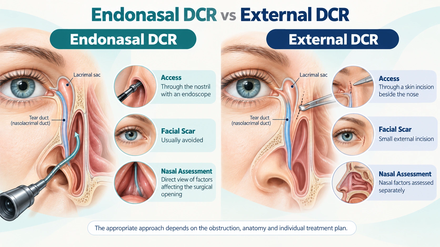
Endonasal DCR and External DCR Compared
Neither technique is automatically better for every patient. Previous surgery, anatomy, level of obstruction and the condition of the tear canal guide selection.
Evaluation Before Endoscopic DCR
The history helps distinguish drainage blockage from dry-eye irritation, allergy and eyelid problems that can also cause watering. The tear drainage system may be assessed by an eye specialist through syringing or other tests. Diagnostic nasal endoscopy allows the ENT surgeon to examine the septum, turbinates, nasal lining and intended surgical area. Imaging is reserved for selected cases, such as trauma, mass, unusual swelling or revision surgery.
Active nasal allergy, sinus disease or infection may need treatment before or alongside the procedure. Blood-thinning medicines and general medical conditions should be discussed during preoperative planning.
What Happens During Endonasal DCR?
The procedure is generally performed under anaesthesia. An endoscope is passed through the nostril, and a small area of bone over the tear sac is opened. The surgeon creates a direct pathway between the sac and nasal cavity so tears can bypass the blocked nasolacrimal duct. A temporary silicone stent may be placed in selected patients to support healing.
The operation does not reduce normal tear production. It aims to restore drainage so tears no longer overflow or remain trapped in an infected sac.
Recovery and Nasal Care
Mild blood-stained nasal discharge, congestion and discomfort can occur initially. Patients should avoid nose blowing, heavy lifting and disturbing the inner corner of the eye. Prescribed nasal care, eye drops or antibiotics must be used as directed.
Follow-up may include nasal endoscopy to remove crusts and confirm that the new opening remains clear. If a silicone tube was inserted, its removal is planned according to healing. Watering often improves, but temporary fluctuation can occur while swelling settles.
Potential Benefits
- A new pathway for tear drainage
- Reduced overflow onto the cheek
- Fewer tear-sac infections when obstruction is the cause
- No external facial incision with the endonasal approach
Possible Risks
- Bleeding, infection or nasal adhesions
- Scar closure of the new opening
- Persistent watering or need for revision
- Temporary stent displacement when a silicone tube is used
Consult for Endonasal DCR in Indore
Long-standing watery eyes should be evaluated rather than repeatedly treated as conjunctivitis. Patients looking for blocked tear duct treatment or endoscopic DCR in Indore can consult Dr. Abhishek Malviya for nasal assessment and coordinated planning based on the complete tear drainage problem.
FAQ
What does DCR stand for?
DCR means dacryocystorhinostomy, a procedure that creates a new drainage pathway between the tear sac and the nose.
Why does a blocked tear duct cause watering?
Tears cannot drain normally into the nose, so they collect and overflow from the eye.
What is the difference between external and endonasal DCR?
External DCR uses a skin incision beside the nose, while endonasal DCR reaches the tear sac through the nostril using an endoscope.
Will endonasal DCR leave a scar on my face?
The approach is through the nose, so it generally avoids an external facial incision.
Is an ENT surgeon involved in DCR?
An ENT surgeon evaluates and manages the nasal route. Eye and ENT specialists may coordinate care depending on the case.
Is a silicone tube always required?
No. A stent is used selectively based on the obstruction, surgical findings and the treating team’s preference.
When does watering improve?
Many patients improve after surgery, but swelling can cause temporary fluctuation. Follow-up confirms that the new opening remains clear.
Can DCR fail?
Scar tissue can narrow or close the opening. Persistent symptoms are evaluated and revision may be considered when appropriate.
Can both eyes be treated?
Both sides can be affected, but evaluation and timing are individualised according to symptoms and diagnosis.
Where can I consult for endonasal DCR in Indore?
Dr. Abhishek Malviya provides endoscopic nasal evaluation and planning for suitable DCR cases in Indore.
This information is educational. An examination is needed to decide which treatment is appropriate. Benefits, risks and recovery vary between patients.